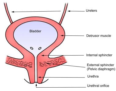
To control the act of urination voluntarily, motor control is achieved through innervation by both sympathetic fibers, most of which arise from the hypogastric plexuses and hypogastric nerves, and parasympathetic fibers, which come from the pelvic splanchnic nerves and the inferior hypogastric plexus. Propel your studies and learn efficiently using the scientifically backed active recall technique! Memorization by reading and re-reading is very ineffective. Surprisingly (or maybe not), a meta-analysis conducted on the effects of different voiding positions in male urodynamics reports that sitting down allows for improved contraction of the detrusor muscle. Whereas GVA fibers on the superior surface of the bladder follow the course of the sympathetic effect nerves back to the CNS, GVA fibers on the inferior portion follow along with the parasympathetic efferent fibers. Sensations from the bladder are transmitted to the central nervous system (CNS) via general visceral afferent fibers (GVA). This signal will encourage the bladder to expel urine through the urethra. The detrusor muscle is a layer of the bladder wall made of smooth muscle fibers that are arranged in spiral, longitudinal, and circular bundles.
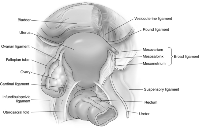
#False ligament of urinary bladder full
There are 2 important pathways involving your bladder: 1) the sensation that lets you know your urinary bladder is full and needs to be voided, and 2) the motor control of your bladder to allow you to urinate at will.įirst, as the bladder walls are stretched when it is full or getting closer to maximum capacity, there are signals that are transmitted through the parasympathetic nervous system to contract the detrusor muscle. The muscles in the bladder that allow for conscious control of when you are or are not in a suitable situation to urinate are especially meaningful in civilized societies. Or to give another mental image, the ureters are posterior to the ovarian/ testicular artery.įor more information about the urinary bladder, take a look below:ĭetrusor urinae muscle, Musculus detrusor urinae With this mnemonic you remember the relationship of these structures. One mnemonic often heard in clinical settings related to the bladder is: “ water (ureters) under the bridge.” This phrase describes an anatomical relationship, between the ureters and the uterine arteries (female) or the ductus deferens (males). During a hysterectomy, where the uterus and uterine artery are removed, the ureter is in danger of being accidentally damaged. While the general volume of the human bladder will vary from person to person, the range of urine that can be held in the bladder is roughly 400 mL (~13.5 oz) to 1000 mL (~34 oz), with the average capacity being 400 to 600 mL.


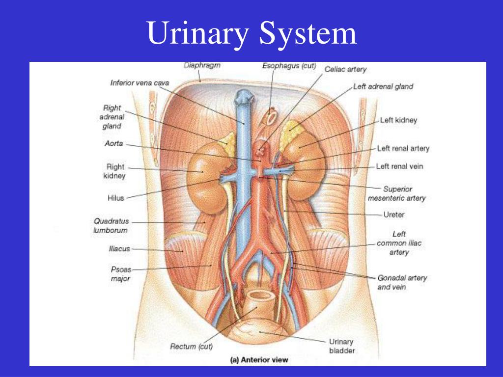
The trigone is the structure that contains the outlet (urethra) of the bladder. The fundus is the base of the bladder, which is formed by the posterior wall and contains the trigone of the bladder, and is lymphatically drained by the external iliac lymph nodes. Urine is collected in the body of the bladder, and finally it is voided through the urethra. It receives urine via the ureters, which are thick tubes running from each kidney down to the superior part of the bladder. Generally, the bladder is a hollow, muscular, and pear-shaped distensible elastic organ that sits on the pelvic floor.


 0 kommentar(er)
